Your shopping cart is empty!
Collagen III alpha 1/COL3A1 Rabbit mAb (20 μl)
| Reactivity: | Human, Mouse, Rat |
| Applications: | WB, IF/IC, IP, ELISA |
| Host Species: | Rabbit |
| Isotype: | IgG |
| Clonality: | Monoclonal antibody |
| Gene Name: | collagen type III alpha 1 chain |
| Gene Symbol: | COL3A1 |
| Synonyms: | EDS4A; EDSVASC; PMGEDSV; Collagen III alpha 1/COL3A1 |
| Gene ID: | 1281 |
| UniProt ID: | P02461 |
| Clone ID: | 7V2P2 |
| Immunogen: | Recombinant fusion protein containing a sequence corresponding to amino acids 150-400 of human Collagen III alpha 1/COL3A1 (P02461). |
| Dilution: | WB 1:1000-1:6000; IF/IC 1:100-1:800 |
| Purification Method: | Affinity purification |
| Concentration: | 0.85 mg/mL |
| Buffer: | PBS with 0.09% Sodium azide, 0.05% BSA, 50% glycerol, pH7.3. |
| Storage: | Store at -20°C. Avoid freeze / thaw cycles. |
| Documents: | Manual-COL3A1 antibody |
Background
This gene encodes the pro-alpha1 chains of type III collagen, a fibrillar collagen that is found in extensible connective tissues such as skin, lung, uterus, intestine and the vascular system, frequently in association with type I collagen. Mutations in this gene are associated with Ehlers-Danlos syndrome type IV, and with aortic and arterial aneurysms.
Images
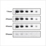
 | Western blot analysis of various lysates using Collagen III alpha 1/COL3A1 Rabbit mAb (A0817) at 1:1000 dilution. Secondary antibody: HRP-conjugated Goat anti-Rabbit IgG (H+L) (AS014) at 1:10000 dilution. Lysates/proteins: 25μg per lane. Blocking buffer: 3% nonfat dry milk in TBST. Detection: ECL Basic Kit (RM00020). Exposure time: 1s. |
 | Western blot analysis of various lysates using Collagen III alpha 1/COL3A1 Rabbit mAb (A0817) at 1:1000 dilution. Secondary antibody: HRP-conjugated Goat anti-Rabbit IgG (H+L) (AS014) at 1:10000 dilution. Lysates/proteins: 25μg per lane. Blocking buffer: 3% nonfat dry milk in TBST. Detection: ECL Basic Kit (RM00020). Exposure time: 60s. |
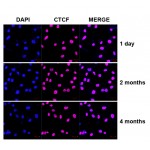
 | Confocal imaging of HeLa cells using Collagen III alpha 1/COL3A1 Rabbit mAb (A0817,dilution 1:100)(Red). The cells were counterstained with α-Tubulin Mouse mAb (AC012,dilution 1:400) (Green). DAPI was used for nuclear staining (blue). Objective: 100x. |
 | Immunoprecipitation analysis of 300 μg extracts from HepG2 cells using 3 μg Collagen III alpha 1/COL3A1 Rabbit mAb (A0817). Western blot was performed from the immunoprecipitate using Collagen III alpha 1/COL3A1 Rabbit mAb (A0817) at a dilution of 1:1000. |
You may also be interested in: